Science up-close: The beautiful and eerie
Take a close-up view of some of science's most intricate and detailed images, part of the 2015 Wellcome Image Awards. Showcasing the best in science imaging talent and techniques, this year's winners include an illustration of pollen grains; a micrograph of a greenfly’s eye; an image showing part of a goat's stomach; plus many more.

An illustration of pollen grains being released from the anther of a flower. This flower is in the Asteraceae family of flowering plants, commonly referred to as the aster, daisy, sunflower, or composite family. The Asteraceae family includes herbs, shrubs, plants and some trees. Pollen is a common trigger of hay fever or seasonal allergies. (Maurizio De Angelis)
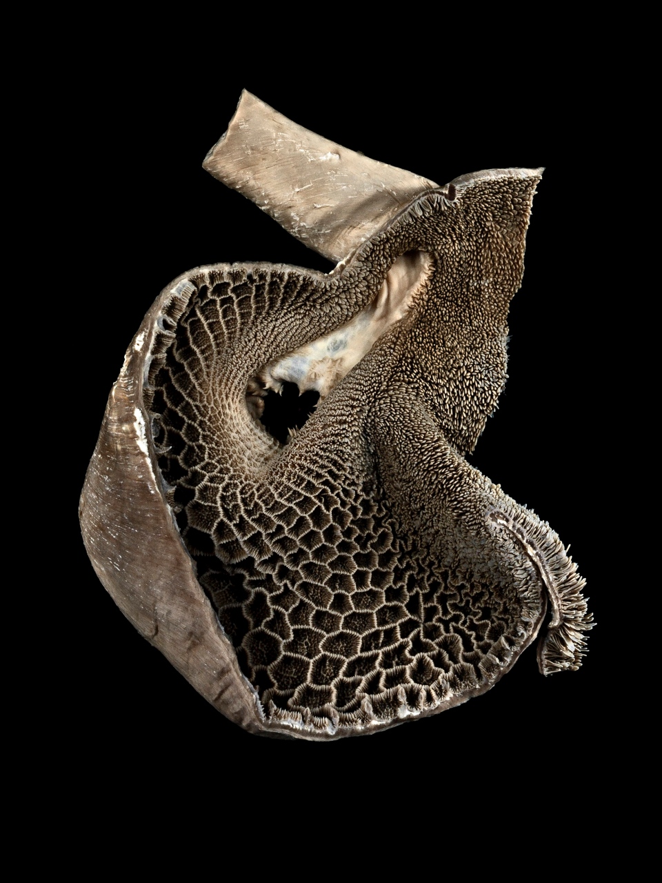
This image shows the reticulum of a goat. The reticulum is one of four chambers in a goat's stomach, and is characterised by the honeycomb pattern which lines the inside. (Michael Frank / Royal Veterinary College)
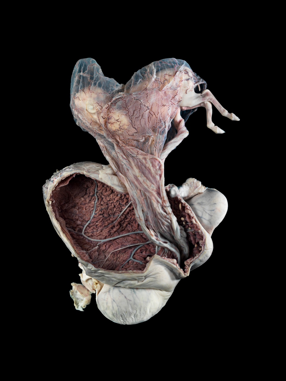
This is a photograph of a mare's (female horse) uterus with the fetus removed but still attached by the fetal membranes and umbilical cord. (Michael Frank / Royal Veterinary College)
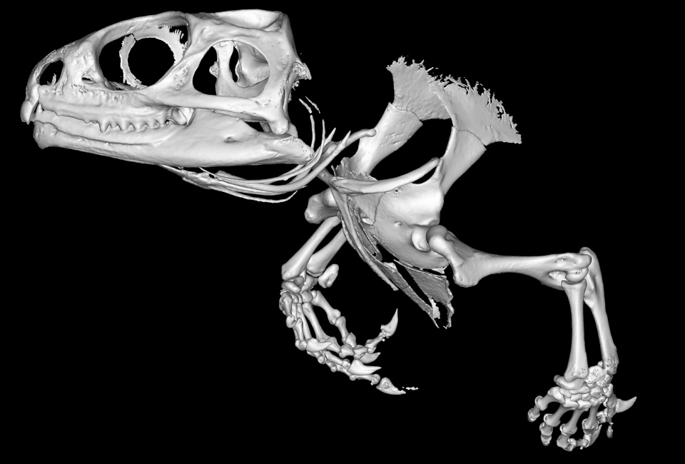
Tuataras are rare reptiles which once shared the Earth with dinosaurs, but are now only found in New Zealand. This is a 3-D reconstruction of the skull and front legs of one. The tuataras are the last member of an ancient group of reptiles, and are sometimes referred to as 'living fossils.' (Sophie Regnault)
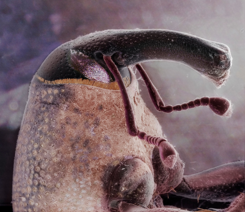
A composite image of the head of a boll weevil. The boll weevil is a beetle which feeds on and lays its eggs in the cotton plant. These agricultural pests have long beaks or snouts and can destroy entire cotton crops. (Daniel Kariko)
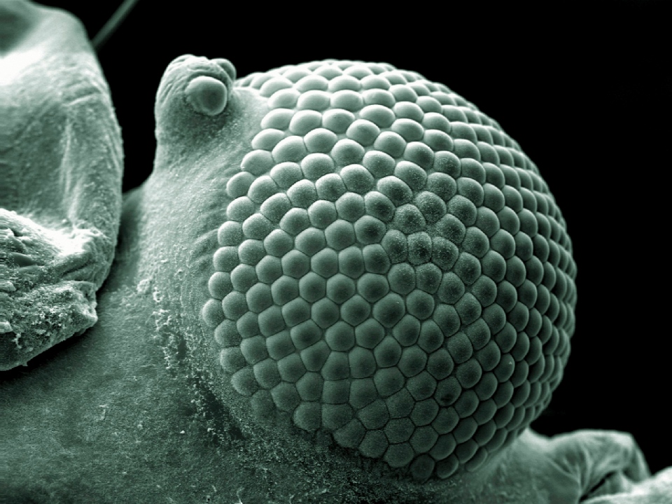
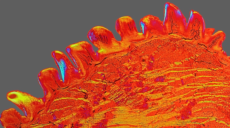
Greenflies have a pair of compound eyes for accurate perception of movement. This scanning electron micrograph shows the insect's eye in great detail. (Kevin Mackenzie / University of Aberdeen)
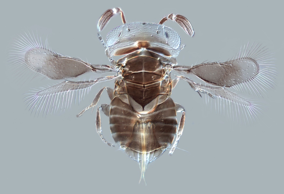
The tiny parasitoid wasp, as viewed from above in a differential interference contrast (DIC) micrograph. The 0.75 millimetre-long wasp belongs to the family Aphelinidae, one of the most effective groups of biological pest control agents. The female wasps lay their eggs in scale insects, which are then killed by the wasp larvae. (Andrew Polaszek / Natural History Museum)
Part of the nervous system in a fruit fly larva is seen in this colourful image. (Albert Cardona / HHMI Janelia Research Campus)
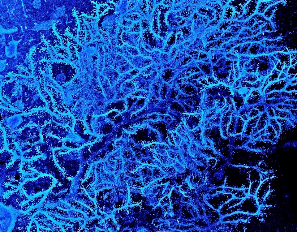
This polarised light micrograph shows a cross section of a cat tongue. This sample is from a vintage prepared slide from the period 1870 - 1905. In a newly-developed technique at that time, blood vessels were injected with dye before fixing and sectioning the tissue in order to visualise the capillaries in the tissue. (David Linstead)
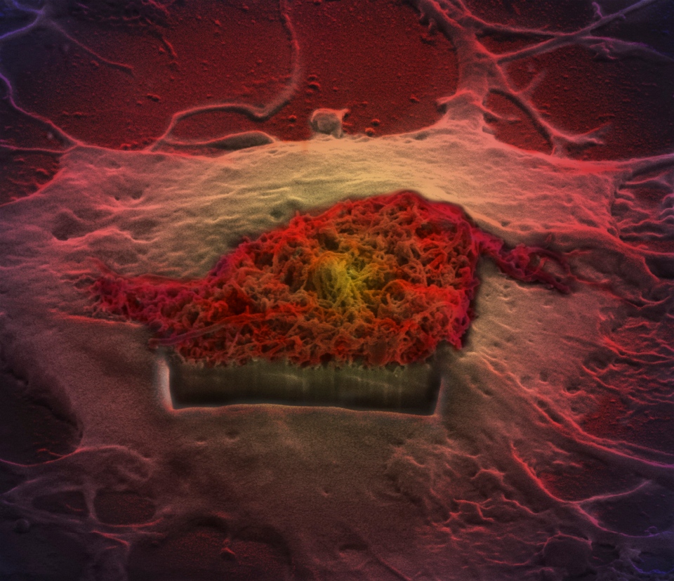
A false-coloured scanning electron microscope showing a brain cell in the process of taking up carbon nanotubes. Carbon nanotubes have been recently explored as drug delivery systems due to their 'nano-needle' characteristics. A section of the cell has been cut to show interactions. (Khuloud T. Al-Jamal, Serene Tay & Michael Cicirko)
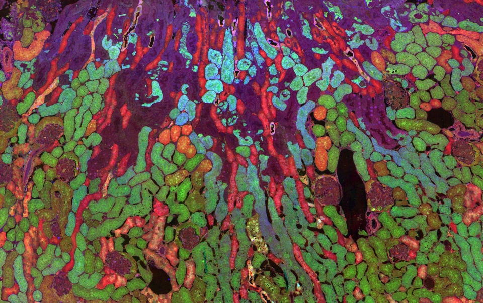
The distribution of metabolites in cells in a mouse kidney is visualized using a technique called computational molecular phenotyping. Metabolites are small molecules produced while chemical reactions take place in a cell to provide energy. (Jefferson R. Brown, Robert E. Marc, Bryan W. Jones, Glen Prusky & Nazia Alam)
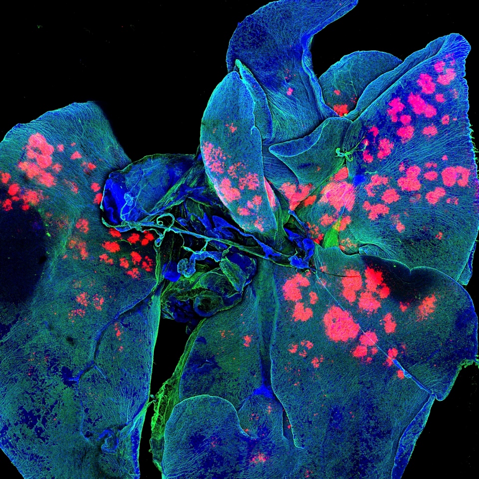
Whole mouse lungs are seen loaded with drug carrying microparticles (red/pink) in this micrograph. The microparticles were also loaded with a fluorescent tracking dye so that they could be visualised one week after administration. (Gregory Szeto, Adelaide Tovar, Jeffrey Wyckoff / Koch Institute / MIT)
An image showing inside the cerebellar cortex of a rat brain, created by a scanning electron micrograph. (Prof. M. Hausser, Sarah Rieubland & Arnd Roth / UCL)
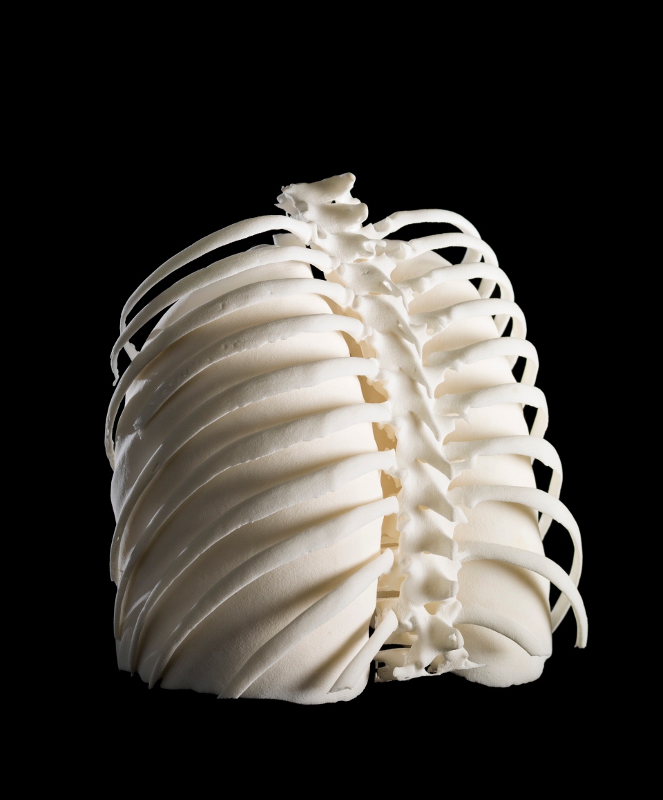
A 3-D printed set of human lungs inside the ribcage, viewed from the back, showing the vertebrae of the spine in the center. The lungs and ribcage belong to a patient who was diagnosed with Hodgkin lymphoma cancer. (Dave Farnham)
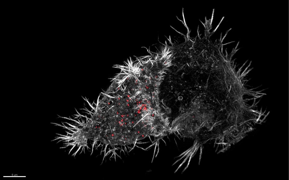
A 3-D structured illumination micrograph of a natural killer cell (left) attacking a susceptible target cell (the less bright, slightly rounder cell on the right). In this image, the natural killer cell has docked onto the target cell creating an area of contact between the two cells. (N. Dieckmann & N. Lawrence)
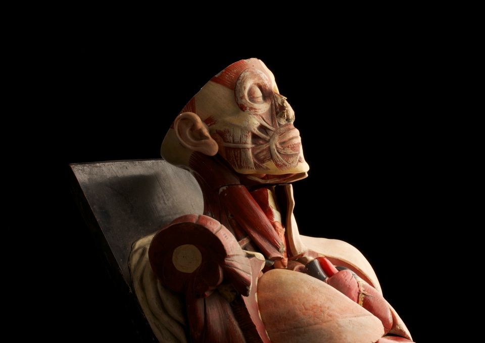
This anatomical model was about to be discarded as garbage when the photographer decided to rescue it and take one last photograph to honour the service it provided to medical students in Trinity College Dublin. (Anthony Edwards)

Designed to provide a distraction for anxious children undergoing painful hospital procedures, this is an interactive multi-sensory unit. The unit is approximately five feet tall and includes a bubble tube, fibre optic lights, mirrors, a solar projector and the capability of producing sound. (Geraldine Thompson / Central Manchester University Hospitals NHS Foundation Trust)

A photo of an elderly woman with Kyphosis, a condition causing curvature of the dorsal spine or 'rounding' of the back and shoulders. This can be congenital or it can be caused by a number of degenerative and endocrine diseases, and is most commonly seen in elderly women. (Mark Bartley / Cambridge University Hospitals NHS Foundation Trust)
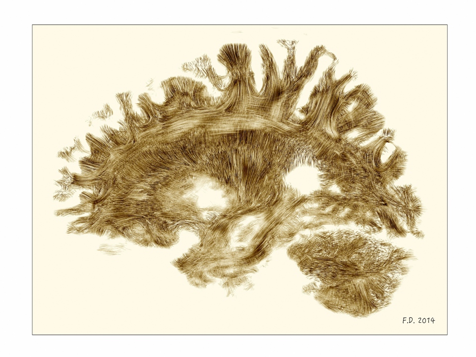
A view of a healthy adult, living human brain, virtually sliced down a vertical axis dividing it into left and right halves. The front of the head is facing towards the left side of the image. (Dr Flavio Dell'Acqua)
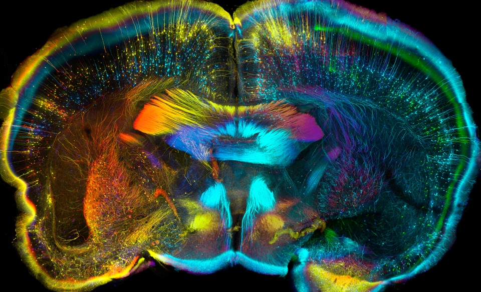
This is a coronal view of a section of mouse brain which has been sliced down a vertical axis to divide it into front and back. (Luis de la Torre-Ubieta / UCLA)
